FEA rupture risk

Finite Element Analysis and Rupture risk

Source: EJVES Mar2015

Finite Element Analysis in Asymptomatic, Symptomatic, and Ruptured Abdominal Aortic Aneurysms: In Search of New Rupture Risk Predictors

This is another interesting addition to the family of predictions for ruptured aneurysm. The strength of it is mainly in its derivation from real life cases and physiologic parameters, ending up in certain immediate outcome (symptomatic, symptomatic, or rupture). And yes, we are more well equipped with info if we have the FEA available with the CTA simultaneously. The problems remains in two things: the lack of adoption of such analysis by surgeons and radiologists (this is the approach of mechanical engineers anyway); and the funding (this article has one declared conflict of interest: one author is the owner/developer of the software he is claiming to work better for us) … yet again, there is no question that the future will ONLY be in such level of analysis, not in the simplified outdated one from the 1990s!!! The new generation has moved to iPhones, Maya software, adobe, smart watches, and smart cars … the technology has doubles 16 (that’s SIXTEEN) times since 1990 (as per Moore law) … so new ideas are now VERY much welcomed!!
here are few interesting findings from the paper: (images are copyrighted to EJVES exclusively). 
1- note how PWRRI is significantly different in 1st vs 3rd group, even after allowing for such large CI. The PWS is also different and less widely distributed; but only for 1st vs 3rd group. 
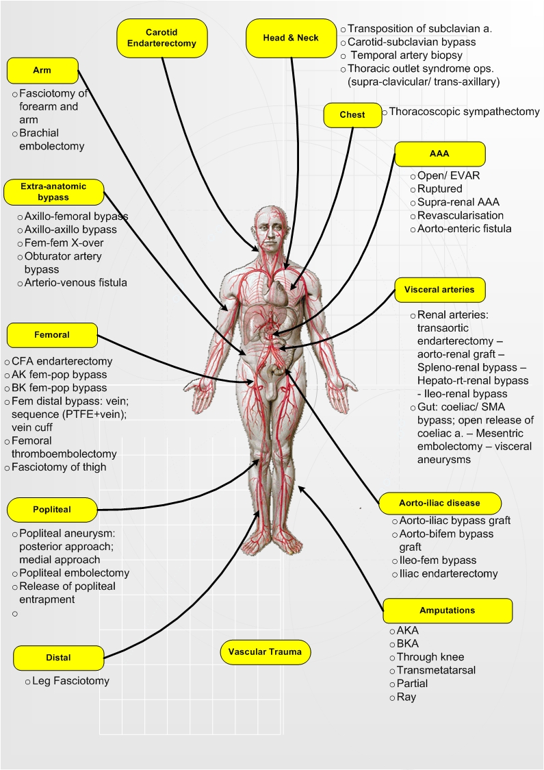
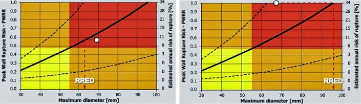
2- Note how in a case of 5.5cm, the PWS can range from 0.1 to 1.0, giving a risk of rupture from minimum to imminent! That’s dangerous indeed!! WHICH PATIENT OF OURS will rupture while we are preparing him/her to undergo a procedure! See my post on estimated risk of rupture, where more specific biomechanical factors (that are calculable by simple mathematics) gives more info on the risk of rupture compared to simple diameter measurements. 
CAR. registry carotid artery aneurysm

Extracranial Carotid Artery Aneurysm (ECAA) registry – use this oppurtunity

ref:

Extracranial Carotid Artery Aneurysm: Optimal Treatment Approach – 

  • J.C. Welleweerd
  • G.J. de Borst
  • on behalf of 
  • the Carotid Aneurysm Registry Project Group
  • EJVES Mar 2015
  • This is an online registry to enter all such rare cases into one database and allow for a proper assessment of best diagnosis and treatment. 
  • I think it is quite useful and certainly attractive to join once one have a case like this!
  • Funding New approaches NICE varicose veins

    Treatment of varicose veins – time for CCG to change their attitude

     ref. NICE Guidelines Aug 2014 – Link

    NICE has made it clear that it is time to change the attitude and offer endothermic/foam/and even surgery for patients who are likely to be struggling with their varicose veins permanently BEFORE going into the compression hosiery option. 

    This is what they stated, and this is what I used recently in a letter to the GP requesting for funding (the outcome of which is not known yet!): 

    Historically surgery and compression therapy were the only treatments available to people with varicose veins, but in recent years other treatments including endothermal ablation and ultrasound-guided foam sclerotherapy have been developed. These newer therapies are less invasive than surgery, promote faster recovery and need shorter hospital stays.

    People with varicose veins are offered treatment with:

    • endothermal ablation (in which the veins are closed off using heat)
    • or, if endothermal ablation is unsuitable, a treatment called ultrasound-guided foam sclerotherapy (in which the veins are closed off using a chemical foam)
    • or, if both endothermal ablation and ultrasound-guided foam sclerotherapy are unsuitable, surgery to remove the varicose veins.

    They should only be offered compression hosiery (stockings designed to improve blood flow by squeezing the legs) as a permanent treatment if none of the other treatments are suitable for them


    bed side teaching vascular curriculum

    Vascular bed-side teaching curriculum – a DGH experience

    It’s important, and interesting, to lead on the quality and quantity of core vascular knowledge that you deliver to trainees (FY1/2, CT1-3, STs) during your ward rounds … regardless of how many ward rounds you do (once weekly; once every six weeks, etc.).

    The following topics and depth of vascular knowledge have to be delivered during your ward rounds; its up to you to design how and over what time you are delivering them.

    1. Basic vascular anatomy and surface anatomy: the tree; origin; branches; access and important structures; essential landmarks 
    2. Basic history and physical examination of the six types of vascular diseases: ICs (with emphasise on risk factors and exercises/smoking); CIL (with emphasis on definitions and correlation to pressures); AAA (with emphasis on acute presentation and decision making); VVs (with emphasis on classification and severity); and diabetic foot (with emphasis on changes in skin, muscles, bones, deformities, nerves, and circulations). 
    3. Core vascular ultrasound scan / waves/ resistance/ eABPIs/ pitfalls/ 
    4. Core pathophysiology – atherosclerosis (stages; endothelium; effect of statin; combination of organ effect) – aneurysmal disease – varicose veins – CILs – diabetic foot – see MRCS presentation 
    5. Core principles of biomechanics in diabetic foot. – see biomechanics presentation
    6. Vascular Facts and Figures 
    7. Basic techniques in vascular surgery: 
    • CORE VIDEOS
    • EXPOSURE: aorta; iliac; femoral; popliteal; foot; carotid; subclavian; axillary; brachial; 
    • PROCEDURES – 
    • LEVEL OF COMPETENCY I would expect from a vascular trainee (with different levels of course): 

    PROCEDURES TO BE DISCUSSED (TO DIFFERENT LEVELS):