complex EVARs radiation

Are teams performing complex EVARs at higher radiation risk over their heads?

ref:

Angulation of the C-Arm During Complex Endovascular Aortic Procedures Increases Radiation Exposure to the Head

EJVES Apr 2015
  • Head dose was significantly higher in the PO compared with the AO (median 54 μSv [range 24–130 μSv] vs. 15 μSv [range 7–43 μSv], respectively; p = .022), 
  • as was over-lead body dose (median 80 μSv [range 37–163 μSv] vs. 32 μSv [range 6–48 μSv], respectively; p = .003). 
  • Corresponding under-lead doses were similar between operators (median 4 μSv [range 1–17 μSv] vs. 1 μSv [range 1–3 μSv], respectively;p = .222). 
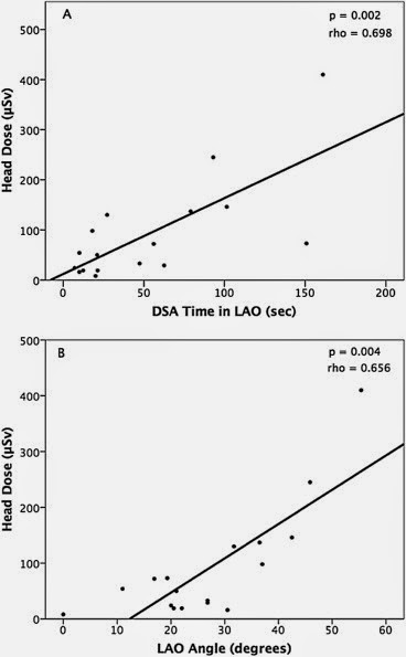
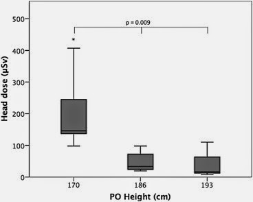
  • Primary operator height, DSA acquisition time in left anterior oblique (LAO) position, and degrees of LAO angulation were independent predictors of PO head dose (p < .05).

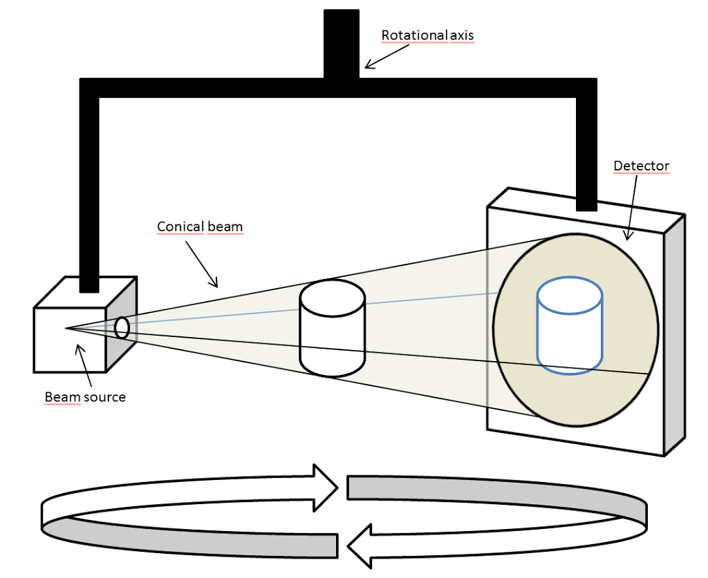
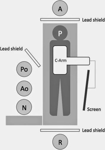
the following diagrams explain it all: 


cone beam CT EVAR

Cone Beam Computed Tomography and EVAR

ref:

Intra-operative Cone Beam Computed Tomography can Help Avoid Reinterventions and Reduce CT Follow up after Infrarenal EVAR

ref: EJVES Apr 2015
Fifty-one patients (44 men) were enrolled in a prospective trial. Patients underwent completion angiography and CBCT during infrarenal EVAR. 

Twelve endoleaks were detected on completion digital subtraction angiography (CA). CBCT detected 4/5 type 1 endoleaks, but only one type 2 endoleak. CTA identified eight type 2 endoleaks and one residual type I endoleak. Two cases of stent compression were seen on CA. CBCT revealed five stent compressions and one kink, which resulted in four intra-operative adjunctive manoeuvres. CTA identified all cases of kinks or compressions that were left untreated. Two of them were corrected later. No additional kinks/compressions were found on CTA. Groin closure consisted of 78 fascia sutures, nine cut downs, and 11 percutaneous sutures. Seven femoral artery pseudoaneurysms (<1 cm) were detected on CTA, but no intervention was needed.

In 1996, three Italian people invented the Cone Beam CT, which went into the US market in 2001. It has become the ‘norm’ in most dental practices, as a simple means of reconstructing the X-Ray easily into 3D. 
 For Interventional Radiology, the patient is positioned offset to the table so that the region of interest is centered in the field of view for the cone beam. A single 200 degree rotation over the region of interest acquires a volumetric data set. The scanning software collects the data and reconstructs it, producing what is termed a digital volume composed of three-dimensionalvoxels of anatomical data that can then be manipulated and visualized with specialized software


atherosclerosis prevention carotid artery disease coronary angiogram

Routine pre-op coronary angio pre- CEA: is it useful?

ref:

Long-term Results of a Randomized Controlled Trial Analyzing the Role of Systematic Pre-operative Coronary Angiography before Elective Carotid Endarterectomy in Patients with Asymptomatic Coronary Artery Disease

EJVES Apr 2015
The group (from Italy) randomised 426 patients who were candidates for CEA, with no history of CAD, a normal electrocardiogram (ECG), and a normal cardiac ultrasound. In group A (n = 216) all patients underwent coronary angiography before CEA. In group B (n = 210) CEA was performed without coronary angiography. Patients were not blinded for relevant assessments during follow-up. Primary end-point was the occurrence of MI at 3.5 years. The secondary end-point was the overall survival rate. Median length of follow-up was 6.2 years.

The outcome is very interesting: short term results are not much different, but long term ones are significantly different and better for coronary patients. This proves the importance of secondary prevention more than anything! 


complex AAA; FEVAR; BEVAR

A propensity-matched comparison of outcomes for fenestrated endovascular aneurysm repair and open surgical repair of complex abdominal aortic aneurysms

Ref.

A propensity-matched comparison of outcomes for fenestrated endovascular aneurysm repair and open surgical repair of complex abdominal aortic aneurysms
EJVES Apr 2015
These figures are shockingly true, and has to be always taken into account when a patient is offered a FEVAR, especially our octogenarians. 
Raux et al.11 compared F-EVAR with open surgery in patients with complex AAA in two large volume centres: In this study, 
  • F-EVAR had higher 30 day mortality rates (9.5% vs. 2%), 
  • higher procedural complications (24% vs. 7%) 
  • and graft related complications (30% vs. 2%) than open surgery.

The authors conceded that “the learning curve of the endovascular technique was likely to be responsible in part for the adverse outcomes observed in the F-EVAR cohort”

The WINDOWS multicentre French Registry reported outcomes in 268 patients who received F-EVAR or B-EVAR for juxtarenal AAA (group 1), suprarenal AAA and TAAA Type IV (group 2), and TAAA Type I, II, III (group 3).15 In hospital mortality was 6.5% for group 1 patients, 14.3% for group 2, and 21.4% for group 3.

This paper suggests that more complex aortic endovascular procedures (especially branched repair of TAAAs) should be focused within higher volume institutions that can provide all treatment options, have experience in dealing with technically challenging intra-operative problems and where multidisciplinary cooperation is such that the choice of treatment is not influenced by specialty, political, or financial aspects.

personally fully agree .. the amount of complex parameters involved in the treatment of those patients is huge … and require a lot of experience and high level thinking/planning… as well as a relaxed time in the sub-sub-sub specialty in vascular surgery …. for best outcome to be achieved,. 


FEA rupture risk

Finite Element Analysis and Rupture risk

Source: EJVES Mar2015

Finite Element Analysis in Asymptomatic, Symptomatic, and Ruptured Abdominal Aortic Aneurysms: In Search of New Rupture Risk Predictors

This is another interesting addition to the family of predictions for ruptured aneurysm. The strength of it is mainly in its derivation from real life cases and physiologic parameters, ending up in certain immediate outcome (symptomatic, symptomatic, or rupture). And yes, we are more well equipped with info if we have the FEA available with the CTA simultaneously. The problems remains in two things: the lack of adoption of such analysis by surgeons and radiologists (this is the approach of mechanical engineers anyway); and the funding (this article has one declared conflict of interest: one author is the owner/developer of the software he is claiming to work better for us) … yet again, there is no question that the future will ONLY be in such level of analysis, not in the simplified outdated one from the 1990s!!! The new generation has moved to iPhones, Maya software, adobe, smart watches, and smart cars … the technology has doubles 16 (that’s SIXTEEN) times since 1990 (as per Moore law) … so new ideas are now VERY much welcomed!!
here are few interesting findings from the paper: (images are copyrighted to EJVES exclusively). 
1- note how PWRRI is significantly different in 1st vs 3rd group, even after allowing for such large CI. The PWS is also different and less widely distributed; but only for 1st vs 3rd group. 
2- Note how in a case of 5.5cm, the PWS can range from 0.1 to 1.0, giving a risk of rupture from minimum to imminent! That’s dangerous indeed!! WHICH PATIENT OF OURS will rupture while we are preparing him/her to undergo a procedure! See my post on estimated risk of rupture, where more specific biomechanical factors (that are calculable by simple mathematics) gives more info on the risk of rupture compared to simple diameter measurements. 
CAR. registry carotid artery aneurysm

Extracranial Carotid Artery Aneurysm (ECAA) registry – use this oppurtunity

ref:

Extracranial Carotid Artery Aneurysm: Optimal Treatment Approach – 

  • J.C. Welleweerd
  • G.J. de Borst
  • on behalf of 
  • the Carotid Aneurysm Registry Project Group
  • EJVES Mar 2015
  • This is an online registry to enter all such rare cases into one database and allow for a proper assessment of best diagnosis and treatment. 
  • I think it is quite useful and certainly attractive to join once one have a case like this!
  • Funding New approaches NICE varicose veins

    Treatment of varicose veins – time for CCG to change their attitude

     ref. NICE Guidelines Aug 2014 – Link

    NICE has made it clear that it is time to change the attitude and offer endothermic/foam/and even surgery for patients who are likely to be struggling with their varicose veins permanently BEFORE going into the compression hosiery option. 

    This is what they stated, and this is what I used recently in a letter to the GP requesting for funding (the outcome of which is not known yet!): 

    Historically surgery and compression therapy were the only treatments available to people with varicose veins, but in recent years other treatments including endothermal ablation and ultrasound-guided foam sclerotherapy have been developed. These newer therapies are less invasive than surgery, promote faster recovery and need shorter hospital stays.

    People with varicose veins are offered treatment with:

    • endothermal ablation (in which the veins are closed off using heat)
    • or, if endothermal ablation is unsuitable, a treatment called ultrasound-guided foam sclerotherapy (in which the veins are closed off using a chemical foam)
    • or, if both endothermal ablation and ultrasound-guided foam sclerotherapy are unsuitable, surgery to remove the varicose veins.

    They should only be offered compression hosiery (stockings designed to improve blood flow by squeezing the legs) as a permanent treatment if none of the other treatments are suitable for them


    bed side teaching vascular curriculum

    Vascular bed-side teaching curriculum – a DGH experience

    It’s important, and interesting, to lead on the quality and quantity of core vascular knowledge that you deliver to trainees (FY1/2, CT1-3, STs) during your ward rounds … regardless of how many ward rounds you do (once weekly; once every six weeks, etc.).

    The following topics and depth of vascular knowledge have to be delivered during your ward rounds; its up to you to design how and over what time you are delivering them.

    1. Basic vascular anatomy and surface anatomy: the tree; origin; branches; access and important structures; essential landmarks 
    2. Basic history and physical examination of the six types of vascular diseases: ICs (with emphasise on risk factors and exercises/smoking); CIL (with emphasis on definitions and correlation to pressures); AAA (with emphasis on acute presentation and decision making); VVs (with emphasis on classification and severity); and diabetic foot (with emphasis on changes in skin, muscles, bones, deformities, nerves, and circulations). 
    3. Core vascular ultrasound scan / waves/ resistance/ eABPIs/ pitfalls/ 
    4. Core pathophysiology – atherosclerosis (stages; endothelium; effect of statin; combination of organ effect) – aneurysmal disease – varicose veins – CILs – diabetic foot – see MRCS presentation 
    5. Core principles of biomechanics in diabetic foot. – see biomechanics presentation
    6. Vascular Facts and Figures 
    7. Basic techniques in vascular surgery: 
    • CORE VIDEOS
    • EXPOSURE: aorta; iliac; femoral; popliteal; foot; carotid; subclavian; axillary; brachial; 
    • PROCEDURES – 
    • LEVEL OF COMPETENCY I would expect from a vascular trainee (with different levels of course): 

    PROCEDURES TO BE DISCUSSED (TO DIFFERENT LEVELS): 

    Posts pagination