Endoleak EVAR graft Uncategorized

Chimneys and periscopes are good enough … 

Ref. EJVES MAY 2916

This is a series of a 100 cases, with total 224 chimney/periscope devices used. The outcome are fairly good as follows:

CPG immediate technical success was 99% (222/224 branches). Mean follow up was 29 months (range 0–65; SD 17); 59% patients were followed > 2 years, 30% > 3 years, and 16% > 4 years. Post-operatively, CPG occlusion was observed early (≤30 days) in three (1.3%) branches and during follow up in 10 (4.5%). At 36 and 48 months, the estimated primary patency was 93% and 93%. After corrective percutaneous (10) or surgical (3) re-interventions, the estimated secondary patency was 96% and 96%. Thirty day mortality was 2%; at 36 and 48 months the estimated patient survival was 79%. Significant shrinkage (72 [SD 23] vs. 62 [SD 24] mm; p < .001) was observed, with a substantial reduction (>5 mm) in 55 patients, and sac enlargement in four. Incomplete aneurysm sac sealing was treated successfully by a secondary intervention in 15 patients.

AAA Embolisation Endoleak EVAR Uncategorized

Amplatz embolisation of internal iliac artery in preparation for EVAR. the biggest series ever! 

Source: EJVES MAY 2016

This is a multi centre series of 169 patients who neededamplatz to proceed with their surgery. Out of all, technical success rate was 97.6%. The embolisation was uni or bilateral. Buttock claudications occurred in ~ 25%, almost 60% of them resolved at first follow up. Bowel is genera occurred in 2 patients (1%) and east treated with limited bowel resection. 

AAA Endoleak EVAR

Is it possible to predict and protect against endo leak type II

Ref EJVES MAY 2016

The answer is yes, if we look at this case control study. Identifying  6 afferent vessels, or the presence of AAA thrombus volume ratio (VR%) <40% have been shown clearly and significantly to be asociatied with increas type II risk rate, and that emboli station of which has reduced significantly the type II endoleak when compared to a arching group 30% vs 75% at 6 months and similar at 12 months. 

So it is so correct to presume this is a first step towards reducing the annoying type II endoleak risk. 

carotid artery disease carotid disease Uncategorized

Carotid plaque echolucency is simply dangerous

Ref. EJVES MAY 2016

A retrospective analysis of well collected data in ACST1 trial, looking specifically on the presence of clear echolucency (homogenous > 25%) vs clear nonecholuecency shows a significant different in stroke rateb8% vs 3%. The risk of ten presence of echolucency cry increases therefore by 2.5 times. 

This is now significant level of evidence, and has to be taken always into consideration. 

Radiofrequency Ablation RFA Venus eczema

New Venous reporting system addressed by the CEO

The new ASPVCS venous reporting system has been addressed by the Ashford and St Peter’s Hospital Trust’s CEO, Suzanne Rankin.


Week ending Friday 22nd April 2016

A message to all staff from Suzanne Rankin,
Chief Executive


Next month we will be celebrating our Staff Achievement Awards which is one of my absolute favourite ASPH events – our very own Oscars!  Congratulations to all our finalists – announced this week on Aspire – and best of luck in the final awards.


Thinking about the awards reminds me of how much we have to celebrate.  While our working environment continues to be tough, every day colleagues across our hospitals are doing fantastic things to improve care for our patients, often going well beyond that ‘extra mile’.  But what we often don’t do so well is promoting this success and I for one would like to see more of that!


Just last week, Valerie attended the Institute for Healthcare Improvement conference along with colleagues Dr Radcliffe Lisk, Dr Keefai Yeong, Dr Clarence Chikusu and Mark Hinchcliffe.  What they didn’t know was that one of our Junior Doctor colleagues, Joe Gallagher, was also there to present a poster highlighting work he had done on the emergency care pathway in a previous Trust.  What a great achievement – well done Joe!  Keefai also had a poster accepted on the work he is doing with junior doctors on improving patient safety – another achievement for Team ASPH!


Talking this through later in the week, we know there are lots of colleagues who are doing similar things, presenting at conferences, having articles published, undertaking research and so on – and we should be celebrating this.  So from now on, I would like to invite you to send us more of your success stories – don’t hide away, let everyone know how well you’re doing.  In a similar way to our weekly round-up of patient feedback, we’re going to start a regular column in Aspire dedicated to your achievements.  And they don’t all have to be about national conferences and academic publications – whatever you’ve achieved, we’d love to hear about it.  We’d also like to see more stories in the local press – service developments, new pieces of equipment or ways of working – whatever it is, let us know.


So I was really pleased earlier this week when I heard from Consultant Vascular Surgeon, Mr Abdullah Jibawi, who along with a number of colleagues, has developed an innovative classification system for venous diseases that can be used during various types of varicose vein surgery.  This is a really innovative piece of work, as nothing like this has existed before, and has been named the Ashford and St Peter’s Venous Classification system (ASPVCS) after our Trust.  It has also been recognised in a number of national conferences.  What a great piece of work!


Please send your stories to the team at comms@asph.nhs.uk or alternatively just drop me a line.  Your achievements are what makes Team ASPH great and we should make more effort to celebrate that whenever we can.


We were also shortlisted recently in this year’s national CHSK Top Hospitals Awards for Quality of Care to patients – an award we already hold from 2015!  We’ll know if we’ve won the award again later next month and if we do, we’ll be sure to celebrate it with you.


In the meantime, enjoy the weekend whatever you are doing.


With very best wishes,


Suzanne Rankin
Chief Executive


Radiofrequency Ablation RFA varicose veins

Introducing the Ashford & St Peter’s Venous Classification System (ASPVCS)

The Ashford & St Peter’s Venous Classification System (ASPVCS) is a new reporting system developed at Ashford and St Peter’s Hospitals NHS Foundation Trust by the vascular team. There has been no reporting system in the scientific literature to quantify and report anatomical complexity that surgeons face while performing varicose vein minimally invasive surgery such as radiofrequency or laser therapy. This lack of standardisation of reporting has marked the varicose vein surgery despite the high number of procedures performed each year in the NHS (over 24700 cases in 2012-13 [1] ).

ASPVCS is constructed using four domains: number of truncal veins treated, number of zone avulsions, number of major anatomical variations (e.g. significant bending), and number of minor anatomical variation (e.g. need for side pressure). ASPVCS classification was found to correlate well with total number of main vein trunks and number of major anatomical variations. ASPVCS scoring was presented at the Association of Surgeons in Training (ASiT) conference 2016. [2]

ABSTRACT:

Association of Surgeons in Training Conference 2016

Abstract: 627;  Evaluating a new intraoperative classification system for reporting complexity level in endovenous procedures – the ASPVCS classification

Objective: to construct new classification for reporting complexity in endovenous procedures -the Ashford & St Peter’s Venous Classification System (ASPVCS).

Method – ASPVCS is constructed using four domains: number of truncal veins treated, number of zone avulsions, number of major anatomical variations (e.g. significant bending), and number of minor anatomical variation (e.g. need for side pressure). Total operative duration used as proxy for level of complexity. Effect of each domain on duration quantified using correlation and Regression analysis.

Results – ASPVCS classification applied on 69 patients undergoing 82 procedures. Median age was 64. Number of main truncal veins treated was 1 (55%), 2 (25%), 3 (16%) and 4 (4%). Major anatomical variations found in 45% of cases. Average procedure duration was 44 min (13-155 min). Significant correlation found between operative duration and total number of main vein trunks (0.62. p<.0001) and number of major anatomical variations (0.36. p < 0.05). Multiple regression analysis showed all domains apart from minor anatomy variation do explain variance in operative duration (R2 = .55, R2Adjusted = .52, p < .05).

Conclusion – ASPVCS scoring can be used in reporting and predicating outcome for intraoperative anatomical variation and is correlated to level of procedure complexity.

References[edit]

  1. Jump up ^ “Vein surgery in the UK”. Retrieved 27 October 2013.
  2. Jump up ^ “ASiT Conference 2016”.
AAA AAA SCORE aortic surgery atherosclerosis prevention bypass carotid artery aneurysm carotid artery disease carotid disease claudication; bypass; angioplasty; complex AAA; FEVAR; BEVAR complex EVARs coronary angiogram critical ischaemia EVAR Health and fitness rupture risk ruptured aneurysm smoking Thoracic aneurysm vascular curriculum

Vascore – an intuitive tool to capture patients’ risk for arterial surgery

apple-app-store

iTunesArtwork Vascore – 

An all-in-one app to check your patient’s suitability for a vascular procedure, calculate the cardiac risk, kidney injury risk, and predict mortality risk based on simple tick boxing exercise. The app provides a tool to calculate the maximum AAA wall stress, allowing for predicting the risk of rupture as well. All results can be easily and simply sent with a one click using secure email (such as nhs.net) where necessary.

graft

Infected Aortic graft – what to do, and what are the outcomes?

ref. EJVES Feb 2016

This one centre long series (55 cases) shows a very good outcome relative to the huge problem that we face with infected graft.

Overall Kaplan–Meier survival was 90.7% at 30 days, 81.5% at 1 year, and 59.3% at 5 years. Graft rupture occurred in three (5%) cases, two of which were caused by graft re-infection (4%). Four patients required major amputation, one of them on arrival and three (5%) during the post-operative period. Nine (16%) patients needed interventions for the vein graft, and two graft limbs occluded during follow up.

Operative tips and tricks

For NAIS, FVs were harvested from popliteal fossa to femoral confluence, leaving the deep FV patent for venous drainage. Side branches were ligated and secured with clips. The first 20 grafts were used in a reversed manner; later on veins were everted and valves excised under direct vision. In most cases, grafts were split proximally and sewn together to match the aortic diameter in a “pantaloon” configuration with 4-0 polypropylene suture. Simultaneously, an infected graft was exposed by a midline (n = 44) or thoraco-lumbar incision (n = 11) and anastomoses were identified. After heparinization the aorta and graft limbs were clamped, grafts were excised, and clearly infected tissue was debrided. The proximal anastomosis of NAIS was reinforced with a piece of tensor fascia lata ( Figure 1 and Figure 2). If there was obvious pus present, the tunnels were flushed with hydrogen peroxide and saline before a new graft was inserted. In a vast majority of cases, distal anastomoses were performed in the groin and covered with a sartorius myoplasty. In a few cases of graft limb shortage, the limb was continued with a saphenous spiral graft or the anastomosis was done more proximally to the iliac artery while closing the femoral artery with a venous patch. When AEF was present, intestinal resection or suture repair was performed by a gastric surgeon. The NAIS was covered with retroperitoneal tissue or omentoplasty and drains were left into the abdominal cavity and to both sartorius muscle pockets.

Uncategorized

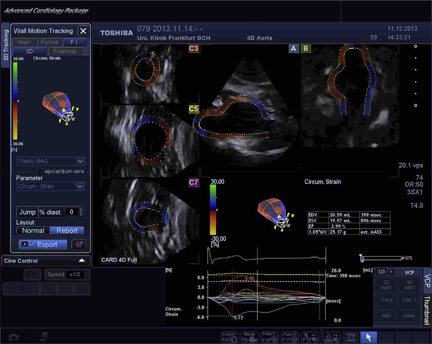
New Vascular Ultrasound Technologies can change our AAA risk assessment

ref. EJVES Feb 2016

High Resolution Strain Analysis Comparing Aorta and Abdominal Aortic Aneurysm with Real Time Three Dimensional Speckle Tracking Ultrasound

This new technology is so promising .. as measuring wall stress is the ONLY way to reliably and easily assess the risk of rupture; the 5.5cm threshold is too crude to be used for the future.

4D USS. For demonstration purposes only. The picture is copyrighted to the publisher.
carotid artery disease carotid disease

Is Carotid intraoperative shunting a harmful procedure?

ref EJVES Feb 2016

The answer is: VERY LIKELY; In one of the largest reported analysing subclinical cerebral changes associated with CEA, it was demonstrates that awake patients who need a shunt are more prone to develop diffusion-weighted magnetic resonance imaging brain scans (DWI) lesions detected by MRI scans 5 days later, and the use of a shunt is an independent predictor.

One in every eight CEA patients developed new DWI lesions (rate doubled in symptomatic patients). Shunt dependence in conscious CEA patients is highly associated with the development of new DWI lesions compared with non-shunted patients.


see also the comments on this article

Posts navigation